Introduction to Animal and Plant Cells

Animal & plant cell coloring worksheet – Embark on a journey of discovery into the microscopic world, where the building blocks of life—cells—reveal their intricate designs. Just as a magnificent cathedral is constructed from countless bricks, so too are all living organisms composed of these fundamental units. Understanding the structure and function of cells is akin to understanding the very essence of life itself; it is a path to enlightenment in the grand tapestry of creation.Animal and plant cells, while sharing some fundamental similarities, exhibit key differences that reflect their distinct roles in the natural world.
These differences, though subtle at first glance, are crucial in determining the unique characteristics and functions of each organism. Consider the mighty oak tree, its strength and resilience, compared to the graceful flight of a hummingbird—each a testament to the cellular marvels within.
Key Organelles and Their Functions
The cell, a microcosm of complexity, houses various specialized compartments called organelles. Each organelle performs specific tasks, contributing to the overall health and function of the cell, much like the specialized workers in a bustling city each contributing to the city’s overall prosperity. Understanding these organelles and their roles provides a deeper appreciation for the elegant design of life.
While animal and plant cell coloring worksheets offer a valuable educational tool for understanding cellular structures, it’s important to remember that learning should be engaging. A break from the intricacies of organelles might be beneficial; consider supplementing your studies with some delightful visuals, such as the charming selection available at cute baby animals coloring pages , before returning to the more complex diagrams of plant and animal cells.
This approach ensures a more balanced and enjoyable learning experience.
Both animal and plant cells possess several common organelles: the nucleus, the control center containing the genetic blueprint; the mitochondria, the powerhouses generating energy; the ribosomes, the protein factories; and the endoplasmic reticulum and Golgi apparatus, involved in protein modification and transport. These organelles, working in harmony, ensure the cell’s survival and proper functioning.
Distinguishing Features of Plant Cells
Plant cells distinguish themselves from animal cells through the presence of several unique organelles. The most notable is the chloroplast, the site of photosynthesis, where sunlight is harnessed to convert carbon dioxide and water into energy-rich sugars—a process that sustains much of life on Earth. This is akin to a solar panel within the cell, capturing the sun’s energy for the plant’s use.
The rigid cell wall, composed of cellulose, provides structural support and protection, giving plants their characteristic firmness and shape. Finally, large vacuoles serve as storage compartments for water, nutrients, and waste products, contributing to the plant’s overall turgor pressure and maintaining its shape.
Significance of Understanding Cellular Structures
The study of cellular structures is not merely an academic exercise; it holds immense practical significance. Understanding cellular processes is crucial in various fields, from medicine to agriculture. For example, advancements in our understanding of cell biology have led to breakthroughs in disease treatment, including targeted drug therapies and gene editing technologies. Similarly, knowledge of plant cell structure allows us to develop improved crop varieties that are more resilient to diseases, pests, and environmental stresses, thereby enhancing food security.
This knowledge empowers us to care for God’s creation in a more informed and effective way.
Worksheet Design and Structure: Animal & Plant Cell Coloring Worksheet
Let us embark on a journey of creation, crafting a learning tool that will illuminate the wonders of the microscopic world within each of us. Just as a skilled artisan carefully chooses their materials, we will thoughtfully design a coloring worksheet that fosters understanding and joy in learning about animal and plant cells. This worksheet will not merely be a coloring activity; it will be a pathway to deeper comprehension, a testament to the beauty of creation.Our goal is to create a worksheet that is both engaging and educational, nurturing a child’s innate curiosity and reverence for the intricate design of life.
Remember, every stroke of color is a step towards understanding the divine artistry of nature.
Layout and Organelle Labeling
The layout should be visually appealing and uncluttered, suitable for the focus and attention span of elementary school students. A large, central image of both an animal and a plant cell will serve as the foundation. Each organelle should be clearly Artikeld and labeled with simple, easy-to-understand terms. For instance, the nucleus could be labeled “The Control Center,” the mitochondria “The Powerhouse,” and the cell wall (for the plant cell) “The Protective Wall.” Use bright, contrasting colors to make the labels stand out against the cell diagrams.
Consider using a simple, child-friendly font. The layout should promote a sense of calm and order, mirroring the harmony found within the cells themselves.
Visual Representation for Understanding
Visual learning is a powerful tool, especially for young minds. The worksheet should leverage this by using vibrant colors to differentiate organelles. The mitochondria, for example, could be colored a bright red to represent their energy-producing function. The nucleus, a central control center, might be depicted in a calming blue. The use of color not only makes the worksheet more engaging but also aids memory retention.
This approach helps students associate specific functions with specific visual cues, creating a strong mental connection between form and function. The visual harmony will encourage a peaceful learning experience.
Organization for Optimal Learning
The worksheet should be structured logically, progressing from simpler concepts to more complex ones. The labeling of organelles should be clear and concise, avoiding overwhelming the child with too much information at once. Perhaps, a small space could be provided next to each labeled organelle for students to write a brief description of its function in their own words.
This encourages active participation and deeper understanding, transforming passive coloring into active learning. This structured approach ensures that the learning process is smooth and enjoyable, mirroring the natural flow of life itself.
Organelle Focus
Embark on this journey of cellular discovery with a spirit of wonder and reverence for the intricate design of life. Just as a magnificent cathedral reveals the dedication of its builders, so too do cells unveil the masterful artistry of creation. Let us delve into the specific components, appreciating their unique contributions to the overall harmony of the cell.
Each organelle within the cell plays a vital role, working in concert to maintain life and sustain growth. Consider each structure a unique instrument in a grand orchestra, each contributing its own distinct sound to create a breathtaking symphony of life.
Cell Wall Structure and Function
The cell wall, a defining feature of plant cells, is a rigid outer layer primarily composed of cellulose. Imagine it as a sturdy fortress, providing protection and structural support for the delicate inner components of the plant cell. This rigid framework maintains the cell’s shape, preventing excessive water uptake and providing resistance against external pressures. The cell wall’s porous nature allows for the passage of water and other small molecules, facilitating communication between cells.
It’s a testament to nature’s ingenuity, providing strength and resilience while maintaining permeability.
Chloroplasts and Photosynthesis
Chloroplasts are the powerhouses of plant cells, the sites where photosynthesis takes place. Picture them as solar panels within the cell, capturing the energy from sunlight. These organelles contain chlorophyll, a green pigment that absorbs light energy, initiating the process of converting light energy, water, and carbon dioxide into glucose (a sugar) and oxygen. On the worksheet, chloroplasts can be depicted as oval-shaped structures containing numerous stacked discs, or thylakoids, which are the sites of light-dependent reactions.
This representation visually communicates their complex internal structure and their essential role in energy conversion. The green color of chloroplasts reflects the presence of chlorophyll, vividly showcasing their photosynthetic function.
Vacuole Function and Size
The vacuole is a large, fluid-filled sac found within both plant and animal cells, though its size and function differ significantly. In plant cells, the vacuole is typically a massive central structure, occupying a significant portion of the cell’s volume. Imagine it as a reservoir, storing water, nutrients, and waste products. This large central vacuole contributes to the cell’s turgor pressure, maintaining its shape and rigidity.
In contrast, animal cells contain smaller, numerous vacuoles, which are involved in various processes, including waste removal and intracellular transport. The difference in size visually reflects the differing roles of the vacuole in plant and animal cells – a large, central storage organelle in plants versus smaller, more numerous vesicles in animals.
Mitochondria Structure and Function
Mitochondria are often referred to as the “powerhouses” of the cell, both plant and animal. These bean-shaped organelles are responsible for cellular respiration, the process of converting glucose into ATP (adenosine triphosphate), the cell’s primary energy currency. Imagine them as tiny energy factories within the cell, constantly working to provide the energy needed for all cellular activities. On the worksheet, mitochondria can be depicted as oval or bean-shaped structures with internal folds called cristae, which increase the surface area for ATP production.
Their presence in both plant and animal cells emphasizes their universal importance in energy metabolism.
Cell Membrane and Cell Wall Differences
The cell membrane and cell wall are both crucial for cell structure and function, yet they differ significantly in their composition and properties. The cell membrane, a feature of both plant and animal cells, is a thin, flexible layer that surrounds the cell’s cytoplasm. It’s selectively permeable, controlling the movement of substances into and out of the cell.
Think of it as a gatekeeper, carefully regulating what enters and exits the cell. The cell wall, present only in plant cells, is a rigid, outer layer made primarily of cellulose. It provides structural support and protection, acting as a strong external barrier. While the cell membrane is dynamic and flexible, the cell wall provides a stable, protective shell.
This difference in rigidity and flexibility reflects their distinct roles in maintaining cell structure and function.
Worksheet Activities and Extensions
Embark on this enriching journey of discovery, where the intricate wonders of animal and plant cells unfold before you. These activities are designed not just to test your knowledge, but to cultivate a deeper appreciation for the miraculous design of life itself. Remember, each cell, a tiny universe, holds a profound story waiting to be unveiled.This section provides interactive exercises and a comparative analysis to solidify your understanding of the fundamental differences and similarities between animal and plant cells.
Consider this a testament to the boundless creativity and ingenuity found within the natural world. Let us delve into the heart of cellular exploration.
Interactive Activities
These interactive activities will help to bring the static images of the worksheet to life, encouraging active learning and deeper comprehension. They offer opportunities for creativity and collaboration, mirroring the collaborative nature of life itself.
- Cell City Analogy: Imagine a cell as a bustling city. Each organelle represents a vital part of the city’s infrastructure. Students can draw a “map” of their chosen cell (animal or plant), assigning roles to each organelle based on its function (e.g., the nucleus as city hall, the mitochondria as power plants, the cell wall as the city walls). This fosters understanding of organelle function through relatable analogies.
- Organelle Matching Game: Create a matching game where students match the names of organelles with their functions and descriptions. This can be done using flashcards or by drawing lines connecting corresponding descriptions. This exercise promotes memory retention and strengthens the association between structure and function.
- Cell Model Creation: Students can create 3D models of animal and plant cells using readily available materials like clay, construction paper, or even recycled materials. This hands-on activity allows for creative expression and deeper understanding of spatial relationships within the cell. The process itself becomes a meditative exploration of form and function.
Comparative Analysis of Animal and Plant Cells
This section encourages critical thinking and observation skills, prompting students to reflect on their coloring and labeling work. It is a chance to appreciate the diversity of life while recognizing the underlying unity of cellular structures.Students should compare and contrast animal and plant cells based on their observations from the coloring exercise. A table can be used to organize their findings, highlighting key differences such as the presence of a cell wall and chloroplasts in plant cells, and the absence of these structures in animal cells.
This exercise encourages detailed observation and analytical skills. Consider the vastness of the natural world reflected in the differences between these seemingly simple structures.
Worksheet Quiz, Animal & plant cell coloring worksheet
This short quiz serves as a gentle assessment of comprehension, a moment of reflection on the journey of learning undertaken. It’s a chance to celebrate the knowledge gained and to identify areas needing further exploration. Approach this not as a test, but as an opportunity for growth.
- What is the function of the cell wall in a plant cell?
- Name three organelles found in both animal and plant cells.
- What is the role of the mitochondria?
- What is the difference between the cytoplasm and the nucleus?
- Why are chloroplasts important for plant cells?
Visual Representation and Table Creation
Embarking on this journey of understanding animal and plant cells is akin to exploring the intricate wonders of creation. Just as a master artist uses diverse techniques to convey a profound message, we too can utilize various methods to grasp the complexities of these microscopic marvels. Let us delve into the visual representation of these cellular structures, using both tables and alternative approaches to deepen our comprehension and appreciation.
Visual aids are essential tools for effective learning. They transform abstract concepts into tangible realities, making complex information more accessible and memorable. By actively engaging with visual representations, we cultivate a deeper understanding and appreciation for the intricate workings of life itself. This process of visualization is a spiritual practice in itself; it allows us to connect with the beauty and order inherent in the natural world, fostering a sense of awe and wonder.
Comparative Table of Animal and Plant Cells
A comparative table offers a structured approach to understanding the similarities and differences between animal and plant cells. This organized presentation allows for a clear and concise comparison, fostering a deeper appreciation for the unique characteristics of each cell type. By carefully examining the data, we can better understand the divine blueprint that underpins all living things.
| Organelle | Animal Cell Description | Plant Cell Description | Key Differences |
|---|---|---|---|
| Cell Membrane | A flexible, selectively permeable outer boundary regulating substance entry and exit. | A flexible, selectively permeable outer boundary regulating substance entry and exit. | Similar structure and function in both. |
| Cytoplasm | The gel-like substance filling the cell, containing organelles. | The gel-like substance filling the cell, containing organelles. | Similar composition and function, though plant cytoplasm often contains more chloroplasts. |
| Nucleus | Contains the cell’s genetic material (DNA), controlling cell activities. | Contains the cell’s genetic material (DNA), controlling cell activities. | Similar structure and function in both. |
| Mitochondria | The “powerhouses” of the cell, generating energy (ATP) through cellular respiration. | The “powerhouses” of the cell, generating energy (ATP) through cellular respiration. | Similar structure and function in both. |
| Ribosomes | Sites of protein synthesis, crucial for cell function. | Sites of protein synthesis, crucial for cell function. | Similar structure and function in both. |
| Endoplasmic Reticulum (ER) | Network of membranes involved in protein and lipid synthesis. | Network of membranes involved in protein and lipid synthesis. | Similar structure and function in both; plant cells may have more extensive ER due to higher metabolic activity. |
| Golgi Apparatus | Processes and packages proteins for transport within or outside the cell. | Processes and packages proteins for transport within or outside the cell. | Similar structure and function in both. |
| Lysosomes | Contain digestive enzymes, breaking down waste and cellular debris. | Less prominent than in animal cells; plant cells rely more on vacuoles for waste breakdown. | Animal cells heavily rely on lysosomes for waste management, while plant cells utilize vacuoles more extensively. |
| Vacuoles | Small, temporary storage sacs. | Large, central vacuole for storage of water, nutrients, and waste products; maintains turgor pressure. | Plant cells have a significantly larger and more prominent central vacuole compared to animal cells. |
| Cell Wall | Absent | Rigid outer layer providing structural support and protection; composed primarily of cellulose. | Presence of a cell wall is a defining characteristic of plant cells, providing structural rigidity. |
| Chloroplasts | Absent | Sites of photosynthesis, converting light energy into chemical energy (glucose). | Presence of chloroplasts is a defining characteristic of plant cells, enabling photosynthesis. |
Alternative Visual Representations
Beyond coloring, consider the transformative power of other visual aids. These alternative approaches can ignite the imagination and deepen understanding, allowing for a more profound connection with the subject matter. Each method offers a unique perspective, illuminating different aspects of cellular structure and function. Consider these as pathways to spiritual growth, mirroring the diversity and richness of God’s creation.
For instance, a 3D model of a cell, meticulously crafted from readily available materials, could offer a tangible and interactive learning experience. Imagine the satisfaction of constructing these miniature worlds, a reflection of the meticulous craftsmanship evident in the natural world. Similarly, a detailed diagram, annotated with clear labels and explanations, provides a comprehensive visual summary of cellular components and their interactions.
Such diagrams serve as a testament to the order and precision inherent in biological systems. A flow chart illustrating the intricate pathways of cellular processes, like photosynthesis or respiration, offers a dynamic visualization of these vital functions. The elegant flow and interconnectedness mirror the harmony and balance found throughout nature.
Illustrative Descriptions for the Worksheet

Embark on this journey of discovery, my friends, as we delve into the intricate beauty of the cellular world. Just as a master artist meticulously crafts a masterpiece, nature has designed the building blocks of life with exquisite detail and purpose. Let us appreciate the divine artistry by visualizing these microscopic wonders. Through careful observation and understanding, we will gain a deeper appreciation for the complexity and elegance of creation.Consider these detailed images as windows into the miraculous world of cells.
Each organelle, like a perfectly placed brushstroke, contributes to the overall harmony and function of the cell, a testament to the grand design of the universe.
Plant Cell Illustration
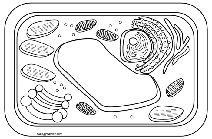
The plant cell, a vibrant testament to life’s ingenuity, typically measures between 10 and 100 micrometers in diameter, exhibiting a rectangular or polygonal shape. Its rigid cell wall, a sturdy outer layer composed primarily of cellulose, provides structural support and protection, much like the strong walls of a castle. Within this protective shell lies the cell membrane, a selectively permeable barrier that regulates the passage of substances into and out of the cell, acting as a wise gatekeeper.
The large, central vacuole, a fluid-filled sac occupying a significant portion of the cell’s volume, maintains turgor pressure, keeping the cell firm and upright, much like a water balloon supporting a structure. The chloroplasts, the powerhouses of photosynthesis, are oval-shaped organelles containing chlorophyll, the green pigment that captures light energy to fuel the process. These are scattered throughout the cytoplasm, like tiny solar panels harnessing the sun’s energy.
The nucleus, the cell’s control center, is a spherical structure containing the genetic material, the blueprints of life, neatly organized and protected. Scattered throughout the cytoplasm are numerous mitochondria, the energy factories of the cell, providing the power for all cellular activities. The endoplasmic reticulum, a network of interconnected membranes, functions as a transport system, moving materials throughout the cell, like an intricate highway system.
Finally, the Golgi apparatus, resembling a stack of flattened sacs, modifies and packages proteins for secretion or use within the cell, acting as a sophisticated postal service.
Animal Cell Illustration
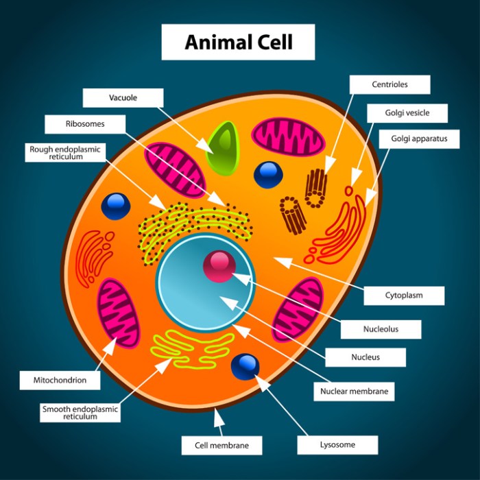
The animal cell, another marvel of biological design, generally ranges from 10 to 30 micrometers in diameter, exhibiting a more rounded or irregular shape compared to its plant counterpart. Lacking a rigid cell wall, it relies on its flexible cell membrane to maintain its shape and regulate the passage of substances. The nucleus, the cell’s command center, is a prominent spherical structure housing the genetic material, the essence of the cell’s identity.
The cytoplasm, a jelly-like substance filling the cell, houses various organelles, each playing a crucial role in cellular processes. The mitochondria, the energy powerhouses, are abundant, supplying the energy needed for the cell’s activities. The endoplasmic reticulum, a network of interconnected membranes, acts as an intracellular transport system, ensuring efficient movement of molecules. The Golgi apparatus, a stack of flattened sacs, modifies and packages proteins, ensuring their proper delivery.
Lysosomes, small spherical organelles, contain digestive enzymes, breaking down waste materials and cellular debris, acting as the cell’s recycling system. The ribosomes, tiny particles scattered throughout the cytoplasm, synthesize proteins, the building blocks of life.
Photosynthesis Illustration
The illustration should depict the process of photosynthesis, where light energy is converted into chemical energy in the form of glucose. Chlorophyll within the chloroplasts captures light energy, which is then used to split water molecules, releasing oxygen as a byproduct. Carbon dioxide from the atmosphere is incorporated into glucose molecules, storing the captured energy. This process, essential for plant life and the overall balance of the Earth’s ecosystem, is a testament to the intricate design of nature.
The image could show a chloroplast with light striking it, leading to the production of glucose and oxygen.
Cellular Respiration Illustration
The illustration should depict the process of cellular respiration, where glucose is broken down to release energy in the form of ATP. The process begins in the cytoplasm with glycolysis, followed by the Krebs cycle and electron transport chain within the mitochondria. Oxygen is consumed, and carbon dioxide and water are released as byproducts. This process, fundamental to all life forms, provides the energy needed for all cellular activities.
The image could show a mitochondrion with glucose entering, and ATP molecules being produced alongside carbon dioxide and water as waste products. The intricate steps of the process could be shown in a simplified, yet informative manner.
